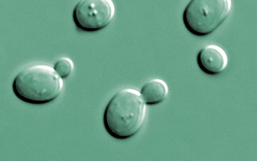
A goal of Stage 1 of our PMM2-CDG PerlQuest with Maggie’s Cure is to identify yeast models that are suitable for use in screens for small molecules that rescue PMM2 deficiency. As described below, we successfully developed screen-ready yeast models that mirror two disease-causing alleles of PMM2-CDG deficiency.
Mutations in the PMM2 gene that produce impaired PMM2 protein with reduced activity cause a form of congenital disorder of glycosylation (CDG). CDG is an inherited metabolic disorder that impairs the production of glycoprotein. PMM2 encodes an enzyme – phosphomannomutase 2 –that catalyzes the conversion of mannose-6-phosphate to mannose-1-phosphate, which is then converted into GDP-mannose. GDP-mannose is a precursor of mannose that is required for protein glycosylation, a form of protein modification where carbohydrates are attached to protein. Deficits in PMM2 enzyme activity limits protein glycosylation, hinders normal cellular function, and affects the nervous system and other organ systems.
SEC53 encodes phosphomannomutase in yeast
The budding yeast Saccharomyces cerevisiae ortholog of PMM2, SEC53, is 55% identical to PMM2. Like PMM2, SEC53 is essential in budding yeast; the loss of SEC53 is lethal. In fact, it’s been known for over 20 years that SEC53 and PMM2 are interchangeable as expression of PMM2 rescues conditional knockout of SEC53 in yeast (Hansen et al., 1997 Glycobiology). This result tells us that we can learn a lot about PMM2 deficiency by studying SEC53 mutants in yeast, an organism that is much more suitable to genetic manipulation and drug screening.
PMM2-CDG is an autosomal recessive disorder where two disease-causing alleles must be inherited from each parent in order for the patient to manifest disease. Three common PMM2 disease-causing alleles are R141H, F119L, and V231M. R141H has no detectable enzymatic activity and never occurs in homozygosity in patients (Kjaergaard et al., 1999 European Journal of Human Genetics). This is consistent with observations in mice, flies and nematodes, where the complete loss of function of PMM2 is lethal.
F119L has 25% enzymatic activity and V231M has 38.5% activity (Kjaergaard et al., 1999 European Journal of Human Genetics; Pirard et al., 1999 FEBS Letters). F119 and V231 are fully conserved in yeast SEC53 (Figure 1; red boxes). Nina, together with our collaborators at Next Interactions, generated the equivalent mutations in yeast. The amino acid positions are offset because the yeast protein is slightly bigger than the human protein: F119L is F126L in yeast; R141H is R148H; and V231M is V238M.
 Figure 1. Sequence alignment of phosphomannomutase genes in human (PMM2) and yeast (SEC53). Asterisk (*) indicates an identical amino acid residue and colon (:) indicates similar amino acids. Red boxes show the conserved disease-causing amino acid residues that we modeled in yeast
Developing the yeast models
Because the mutant variants of PMM2 have low to no detectable enzymatic activity, we decided to express SEC53 at different levels to determine if changes in protein abundance affect the ability of each variant to restore yeast cell growth. To do this, we took advantage of a commonly used genetic tool where we replace one DNA promoter sequence for another (Figure 2). The promoter determines how much mRNA and therefore protein to make from a DNA template. In this case, we placed SEC53 under the control of four different promoters to alter the amount of protein the cell produces over a 40-fold range. You can see in Figure 2 using the green fluorescent protein (GFP) as a readout of protein abundance that we have a range of expression levels relative to the native SEC53 promoter.
 Figure 2. Comparison of promoter strength. Different promoters are used to drive the expression of the gene of interest. GFP is placed under the REV1, SEC53, ACT1, or TEF1 promoter and the fluorescent intensity of GFP is measured by flow cytometry. The graph displays the fluorescence in arbitrary unit (a.u.) against the cell count. The raw numbers are shown in the table along with the expression level relative to the SEC53 promoter
We also had to overcome the complication that SEC53 is an essential gene. In order to generate new SEC53 mutants with little to no detectable enzymatic activity, we employed a strategy where we place a wildtype or fully functional copy of SEC53 on a plasmid that we can conditionally remove. A common tool in yeast is combining a URA3 genetic marker with 5-Fluoroorotic acid (5-FOA). 5-FOA is an analog of uracil that is converted to a toxic compound in cells that synthesize their own uracil, which the URA3 marker enables. Thus, we can place a wildtype SEC53 onto a URA3-marked plasmid and into a cell where the native copy of SEC53 is knocked out (Figure 3).
We can then insert a SEC53 mutant variant, a wildtype copy, or a mock GFP, into the genome of this strain. When we grow these cells in the presence of 5-FOA, there is selective pressure against cells that possess the URA3 plasmid, and only cells that have lost the URA3 plasmid will survive. We can determine whether a SEC53 mutant allele is sufficient for growth and how well it grows compared to wildtype. At this point, we know that wildtype SEC53 will allow normal growth and loss of SEC53 (∆, containing a GFP instead) will cause cell death.
What about yeast cells with the F126L, V238M, or R148H allele?
 Figure 3. Strategy for working with an essential gene in yeast
Characterizing the yeast models
Initial characterization of the strains revealed that the F126L and V238M alleles are sufficient for cell growth. It also showed variability between replicates that I suspected arose from different timing of plasmid loss during the experiment. Therefore, I removed the URA3 plasmid from all promoter combinations with wildtype (WT), F126L, and V238M strains by growing these cells in 5-FOA media (Figure 4, step 1). The SEC53 knockout (∆) and R148H cells require the plasmid carrying a wildtype SEC53 for viability so the plasmid is kept until the beginning of the experiment (Figure 4, step 4). All strains were serial diluted into 5-FOA media and incubated at 30oC to grow. Cell density was recorded at 0, 18, 20, and 24 hours to generate a growth curve.
 Figure 4. Experimental Workflow
Below are representative data from the 20 SEC53 variants grouped by promoter (Figure 5). The far-left panel shows that the V238M allele when expressed at native level is sufficient for growth, but at a slower rate than wildtype. F126L and R148H alleles are not sufficient for growth at the native level. Doubling the expression of V238M alleles with the pACT1 promoter completely rescues this mutant. The F126L allele now becomes sufficient for growth at a reduced rate when its expression is doubled. When we further increase the expression of F126L and V238M alleles to ten-fold above native level with the pTEF1 promoter, these cells are indistinguishable from wild type cells. Together, these data can be fully explained by mass action effects, where a reduction in enzymatic activity can be overcome by increasing the total amount of enzyme.
In the pREV1 chart, I included pSEC53-SEC53 wild type (green) as a control for normal growth in order to determine whether reducing the level of wild type SEC53 compromises growth. As you can see from the far-right panel, reducing the wild type level to 20% of its native level modestly, but consistently, compromises cell growth. This indicates that cells are highly sensitive to the total amount of SEC53 protein.
 Figure 5. Growth of SEC53 variants over time.
Advancing to the drug screening stage
If we now compare in Figure 6 all the strains relative to one another, we can really see that the severity of growth defects of the SEC53 alleles correlate very well with the level of enzymatic activity of the PMM2 alleles. In other words, the genotype-phenotype relationship is conserved between yeast and humans. This is exciting for a couple of reasons. First, a direct relationship between enzymatic activity and physiological phenotype isn’t always observed in biology because the cellular environment is robust and tends to have compensatory pathways. Second, because we can overcome the deficiency of the V238M and F126L variants by increasing their protein abundance, we are even more optimistic that we will find small molecules that can do the same. On the other hand, the R148H allele, which has no detectable enzymatic activity, cannot be rescued by increasing protein abundance even to very high levels.
 Figure 6. Comparison of growth between yeast strains relative to wild type at t = 18 hours
Moving forward, we’ve determined that the pSEC53-V238M and pACT1-F126L alleles are good candidates to advance to the drug screening stage. These models have a growth defect (a phenotype to reverse in a screen) but are not dead (a phenotype too severe to overcome). Our newest Research Associate, Maddie, and I have started pilot screens with our repurposing drug library containing about 2500 compounds. There’s more to come soon!


