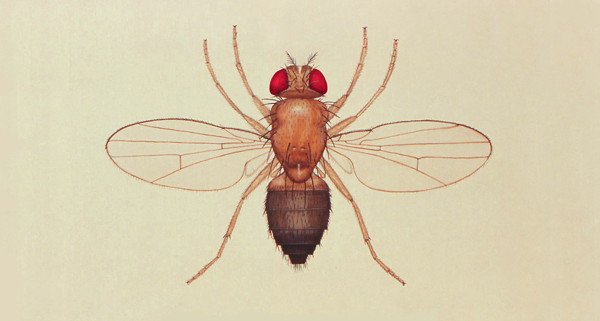
My colleague, Tamy Portillo Rodriguez, and I attended the 59th Annual Drosophila Research Conference – a fantastic meeting of scientists exploring many different fields of biology using the fly – in Philadelphia earlier this month. The conference (in some circles referred to as the ‘Fly Meeting’) is a great opportunity to hear about newly developed tools and methods, learn about exciting disease models, catch up with old friends and colleagues, and generally be exposed to interesting new biology. In this post, Tamy and I will go over some of what we learned at the conference, and the cool work the Drosophila community is pursuing.
Tamy: I’ve had the opportunity to attend the Annual Drosophila Research Conference for three consecutive years, and at each I’ve learned a lot and met great people. This conference was no exception. For example, multiple talks characterized new fly disease models, explored the mechanism of disease biology, or presented new techniques using our amazing fruit flies.
I’ll start by telling you about the work presented by Dr. Eliana Weisz from Perelman School of Medicine at the University of Pennsylvania titled “Drosophila FMRP modulates energy metabolism and mitochondrial function.” Eliana made mutations in fly dfmr1 to model Fragile X syndrome, which is the most common heritable form of intellectual impairment in children, and is also associated with high rate of obesity. At a molecular level, Fragile X syndrome is an X-linked disorder that is caused by a CGG trinucleotide repeat in the FMR1 gene. Eliana used null dfmr1 mutants to identify and characterize the metabolic phenotypes in dfmr1 flies using whole fly tissues. She determined that carbohydrate levels, lipid metabolites, and triglyceride levels were diminished in dfmr1 mutant flies. Moreover, they determined that dfmr1 mutant flies are sensitive to starvation and that mitochondrial morphology is altered in these mutants. This work taught me about several different types of assays that could potentially be used for metabolic studies in flies at Perlara.
Josh: I had the opportunity to meet Dr. Kristi Wharton, Professor of Biology at Brown University, and co-lead a table discussion on Drosophila models of neurological disease with her. Later, she gave a great talk on her work modeling Amyotrophic Lateral Sclerosis (ALS) in flies, “Activation of BMP signaling in non-motor neurons rescues motor dysfunction in a Drosophila model of Amyotrophic Lateral Sclerosis.” ALS is an adult-onset motor neuron disease, and a familial form form of the disorder is caused by mutations in a gene called Superoxide Dismutate 1 (SOD1). Her talk expanded on her previously published work modeling ALS patient mutations in the fly homolog of SOD1, called dSOD1. dSOD1 mutant flies have phenotypes similar to ALS patients; they have movement dysfunction, degeneration of the neuromuscular junction, and die early. Kristi has carefully explored the early cellular mechanisms driving these degenerative effects and made a surprising discovery. First, she found that driving the expression of an important signaling molecule from a protein family called BMPs could rescue some of the locomotor and synaptic phenotypes in these flies. Second, the rescue she observed doesn’t come from BMP expression in motor neurons, but rather in sensory and/or interneurons! This models seems perfect for a drug screen!

Excerpt from Şahin et al. (2017): dSod mutations cause partial pupal lethality and short lifespan in escapers.
Tamy: The second talk that stood out to me was the work presented by Raghuvir Visawanatha from Harvard Medical School titled “Pooled-format, genome-wide CRISPR/Cas9 screening in Drosophila Cells.” Raghuvir and colleagues have resolved the problem of quickly integrating CRISPR guide RNAs (sgRNAs) into insect models using phiC31 system. This allows them to conduct pooled CRISPR screening experiments in a high throughput manner using fly cells. They conducted a genome wide sgRNA pooled CRISPR study on fly cells and were able to identify essential genes with resistance and synergistic interactions. They also demonstrated that phiC31-mediated recombination can be used to deliver complex libraries to fly cell lines. They are currently planning on doing binary genetic interaction screens, making new libraries, Cas9 variants, and alternative selection strategies, as well as expanding the system to other fly and mosquito cell lines so that this new technique can be implemented in other labs. For the Perlara fly team, pooled CRISPR techniques will open doors toward understanding the mechanism of action of our candidate drugs.
Josh: Every year at the Fly Meeting, the Sandler Memorial Award is awarded to a young Drosophila scientist for the best dissertation of the preceding year. The winner this year was Dr. Lucy Liu, for her work in Dr. Hugo Bellen’s lab at Baylor College of Medicine some of which is published here. In it she digs into a common feature of many neurodegenerative diseases, oxidative stress, and links it to a new feature, lipid droplets. Her work follows a series of genetic screens done in the Bellen lab to identify genes that cause photoreceptor degeneration in the fly visual system. Three genes identified in these screens impact mitochondrial biology, sicily, Aats-met, and Marf (fly homologs of NDUFAF6, MFN1/MFN2, and MARS2, respectively), and cause the formation of lipid droplets in cells. Curiously, these droplet aren’t forming in neurons (i.e. the photoreceptors) but rather in surrounding support cells, and they form before any signs of neurodegeneration arise. Moreover, the droplets accumulate in wild-type support cells when only nearby photoreceptors are homozygous mutant. Lucy did some nice work to show that droplet formation is linked to reactive oxygen species (ROS) production. Her hypothesis is that to stave off damage from ROS, neurons are passing oxidized lipids to support cells. This is certainly an interesting idea!

Excerpt from Figure 2. Mutations in mitochondrial genes cause lipid droplets to form in retina support cells (Liu, et al.)
Tamy: Last but not least, I would like to briefly mention the work presented by Nick Matinyan from Baylor College of Medicine and his poster titled “Transgenesis 2.0: Selection-based genome manipulation in Drosophila melanogaster.” Nick used the phiC31 platform to generate transgenic fly lines that have either a drug resistance selection marker or a drug sensitivity counter-selection marker. This allows him to perform a chemical selection on drug treated food to identify transgenic flies with the desired mutation and marker. This essentially is the same principle used in yeast when they are grown on penicillin food when the desired mutant has penicillin resistance on the plasmid construct. For Perlara, this could reduce the time we spend screening our transgenic fly lines, since it would only be a matter of growing flies on the selectable drug food allowing only individuals that carry our transgene-of-interest to survive.
Josh: Overall, it was an exciting and fruitful meeting. We saw some amazing science, reconnected with old friends, came back to Perlara with great new ideas, and met many talented researchers interested in our company. Tamy was tweeting up a storm during the meeting, so check out @Tamy_Portillo on Twitter.
If you are interested in what Perlara’s PerlQuest fly team has been up to, catch up on some of our past posts.
