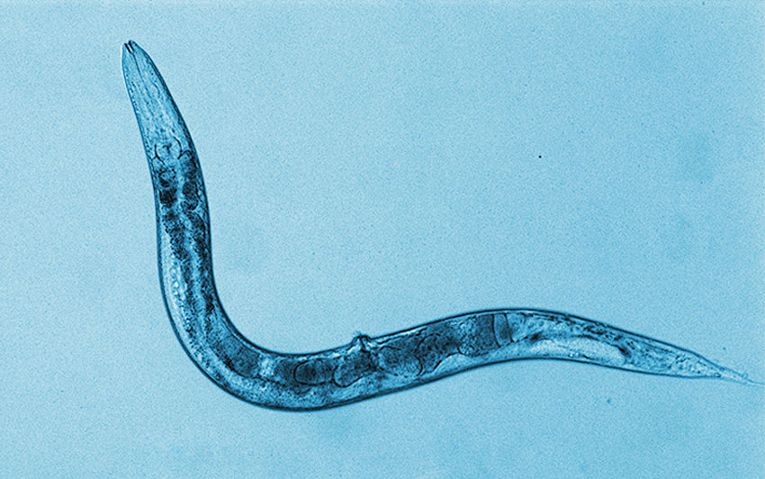
Perlara and Maggie’s Cure have started a PerlQuest for a glycosylation disorder called PMM-2 deficiency disease – one of the most common congenital disorders of glycosylation. Over the past year at Perlara, we have modeled the pmm-2 deficiency disorder in yeast, worms and flies. The post below will describe our efforts in developing a nematode model of PMM-2 deficiency.
PMM-2 deficiency arises from defects in the gene encoding phosphomannomutase-2. Like the name signifies, this enzyme protein is responsible for ‘mutating’ or ‘converting’ a sugar residue on proteins, specifically, the mannose-6-phosphate residue to mannose-1-phosphate. The mannose-1-phosphate tag on proteins is converted to guanosine diphosphate mannose (GDP-mannose). The GDP-mannose tag is required for subsequent conversion to dolichol-P-oligosaccharides. The addition of these sugar moieties at precise locations of complex molecules such as proteins or lipids serves many different functions: proper protein folding, protein stability, protein targeting into appropriate cellular compartments like the endoplasmic reticulum, immunological specifications etc.

Fig.1 Structure of mammalian pmm-2 protein (By Emw – Own work, CC BY-SA 3.0, https://commons.wikimedia.org/w/index.php?curid=8820941) |
Becoming familiar with F52B11.2
F52B11.2 is the gene sequence in C.elegans that has been identified as 54% identical to mammalian PMM-2, but has been virtually unstudied. The nematode pmm-2 protein bears a high degree of similarity with mammalian protein. Importantly, two of the commonly encountered mutant sites, F119 and R141 are conserved in the worm (Fig. 2B). This is noteworthy because we can mutate these sites and determine if the mutant nematode suffers deficits in pmm-2 function like in humans.
| 2A.
2B.
Fig. 2 Nematode pmm-2 gene and protein loci 2A. Representation of nematode pmm-2, grey box indicates 518bp deletion in strain VC3054, pink arrow s indicates orthologous F125L and R147H site that are associated with pmm-2 disease. 2B. Alignment of human protein and nematode protein with mutation sites highlighted in red boxes |
While the function of the gene in nematodes has been virtually unstudied, a knockout model of this gene did exist thanks to the efforts of International C elegans Gene Knockout Consortium (Fig. 2A). The knockout model contains a ~500bp deletion that results in larval lethality, i.e. worms arrest as larvae and eventually die. Unfortunately for worms, which are hermaphrodites and self-fertilize to create progeny, this means that obtaining subsequent generations of worms with the same genotype is impossible.
Exactly how severe is this larval lethality?
3A. 
3B.
Fig. 3 Lifespan of nematodes and progeny created by heterozygous pmm-2+/- strain, VC3054 3A Images of worms through stages of development, top inset shows image of pmm-2 null homozygous worm arrested as larva; bottom inset shows pmm-2 heterozygous animals grown to adulthood in one well of a 384-well plate. 3B. Bars indicate percentage of heterozygous and homozygous pmm-2-/- null animals created across 5 trials. Only 10% of homozygous null animals were observed, these were arrested as larvae. |
Because the homozygous null mutant displays larval lethality, the strain is maintained as a heterozygote on a balancer line. This strain produces only two kinds of progeny- a) heterozygotes bearing one normal copy of pmm-2 and one copy of mutant pmm-2, b) homozygous mutant pmm-2. Heterozygotes bearing a single WT copy of pmm-2 can be distinguished by the presence of a pharyngeal GFP marker. These animals grow to adulthood and reproduce normally. Homozygote nulls lacking pmm-2 are distinguished by lack of GFP. These animals arrest as L1 larvae.
Note: The homozygote progeny carrying two wildtype copies of pmm-2 is non-viable due to chromosomal translocation.
While larval lethality of homozygous null pmm-2 mutants makes for an attractive screenotype for drug discovery, we first determined the percentage of homozygous animals produced per generation. Through experiments, we found that only 10% of the total progeny from the heterozygous animals are homozygous nulls and arrest as larvae (normally we would expect 25%). Because a screen of 360 drugs alone requires ~22000 animals, obtaining enough homozygous animals for a drug discovery screen would be tedious. This challenge led us to create a worm bearing a point mutation in the pmm-2 gene. Thankfully, because the amino acid residues commonly mutated in human pmm-2 disease were conserved in the worm, we generated the F125L (analogous to human F119L) variation in the worm. This mutation is associated with some residual pmm-2 activity in humans, so we reasoned that obtaining a viable worm with some phenotype might be possible.
How does an F125L point mutant worm fare?
Almost too well, actually… To cut a long story involving multiple crosses short, homozygous F125L pmm-2 mutant worms are overtly normal, reproduce normally and have normal growth and developmental timeframes as compared to wildtype worms. We determined using quantitative PCR that the F125L point mutant worm produced nearly the same level of mRNA as a normal or wildtype worm does (Fig.4). So, even if the gene is defective, enough copies of the semi-functional RNA are being generated. Surprisingly, even the heterozygous pmm-2 mutant produced enough copies of the mRNA to match levels in wildtype animals, suggesting compensation from the single normal allele. The lack of any overt phenotypes in the F125L mutant animals meant that we could not use this animal for screening. We did determine that a pmm-2 defect does exist in these worms because when exposed to pmm-2 RNA interference treatment, the F125L animals display abnormal morphological differences compared to wildtypes treated the same way.

Fig 4. Transcript Verification in wildtype and pmm-2 nematode mutantsΔCq plots of transcript levels of pmm-2 amplified using two different primer sets, displayed with reference to reference gene- act-1 in wildtype (N2), F125L pmm-2 homozygous mutants (COP1626 and COP1627) and heterozygous pmm-2 strain (VC3054). |
Stressed for a screen
The lack of an overt phenotype in the F125L point mutants, the apparent compensation from a single copy of pmm-2 in heterozygous animals and the larval lethality in homozygous pmm-2 nulls all converged around one hypothesis- while the complete lack of pmm-2 function leads to lethality of some sort, sub-normal levels of pmm-2 function are sufficient to support normal function and thriving. While preliminary, this finding implies that pmm-2 defects are likely tantalizingly close to some form of rescue. Most humans presenting with the disease suffer from point mutations and not complete enzyme defects i.e. homozygous nulls are unlikely to be walking around us but people with hypomorphic forms of defective pmm-2 are likely walking around us. So, a minor correction to a dysfunctional enzyme is likely to be accompanied by huge corrections in quality of life and thriving. At least, I would predict so…
Fig 5. Example images of bortezomib-exposed wildtype (WT) and pmm-2 F125L homozygotes A) WT and pmm-2 F125L homozygote larvae display sensitivity to 13.6µM bortezomib as early as day 3 of exposure in liquid culture, manifesting as delayed development. B) WT and pmm-2 F125L homozygotes show differences in growth stages achieved after 6 days of exposure to 13.6µM bortezomib. |
How to screen in the absence of a phenotype?
What if we could enhance an underlying pmm-2 defect further by using a chemical that burdens the glycosylation process. We would predict that such a drug would not produce a huge defect in wildtypes because their glycosylation system is intact. But in pmm-2 point mutants, which have a broken glycosylation system, adding an additional stressor would reveal potential deficits. With this in mind, we tested tunicamycin and bortezomib, drugs that have been implicated in proteosomal dysfunction. Tunicamycin has been utilized in worms to replicate glycosylation defects synthetically. However, in our hands, this drug did not differentially affect wildtype and pmm-2 F125L worms. It did produce growth suppression but no differential sensitivity between the strains. Bortezomib, on the other hand, an irreversible proteasome disrupter did produce differential sensitivity between wildtypes and pmm-2 point mutants.
The use of bortezomib to reveal underlying glycosylation defects is not new. Recently, Lerhbach and Ruvkin showed that bortezomib suppression uncovered larval defects in a nematode model of Ngly-1 deficiency. This principle has been utilized for a genetic screen on Ngly-1 deficient worms.
It is interesting that pmm-2 deficient worms which also suffer a defect in the the glycosylation pathway are similarly susceptible to the effects of bortezomib, albeit at a different concentration. Does the differential sensitivity to the same proteosome disrupter underscore differences in the functional significance of the pmm-2 and ngly-1 mutations? We hope to answer this and other interesting questions in our pmm-2 PerlQuest journey. For now, full steam ahead on drug screens for Pmm-2 and NPA worm models.
References:
- Struwe, BW, Hughes, LB, Osborn WD, Boudreau DE, Shaw MDK, Warren EC, ‘Modeling a congenital disorder of glycosylation type 1 in C.elegans:A genome-wide RNAi screen for N-glycosylation-dependent loci’, Glycobiology, 2009; 19 (12):1554-156.
- Lehrbach JN and Ruvkin G, ‘Proteasome dysfunction triggers activation of SKN-1A/Nrf1 by the aspartic protease DDI-1’, eLife, 2016; 5:e17721


