Scientists utilize an array of organisms to gain insight into the mechanisms underlying disease. Among the most commonly used organisms is the fruit fly, Drosophila melanogaster. There are three main reasons that the fly is a terrific organism to study human diseases. First, about 75% of the genes that have the potential to be defective and lead to human diseases also exist in flies. Second, flies are inexpensive to maintain in large populations and have a fast life cycle. Thus, many experiments can be done cheaply in a short period of time. Third, present day Drosophilists have an extremely broad range of tools for manipulating genes to learn about gene function. To date, 6 Nobel Prizes in Physiology or Medicine have been awarded to scientists that primarily used Drosophila in their research. It’s remarkable that nature provided this wonderful model organism, and that Thomas Hunt Morgan, Alfred Sturtevant, Herman Muller and Calvin Bridges recognized its potential over 100 years ago.
Amongst other model organisms, we at Perlstein Lab are using fruit flies to study and find treatments for rare diseases. A previous blog post by Jun Axup described our goal to find therapeutics for patients with alterations in npc1, the primary causal gene in the disorder Niemann-Pick type C. The human and fly Npc1 proteins are similar by 63%. This degree of protein sequence conservation indicates that the human and fly Npc1 proteins have similar or identical functions. In 2005 and 2006, the laboratories of Matthew Scott (Stanford University) and Leo Pallanck (University of Washington) published their descriptions of flies with defective npc1 genes. They correctly reasoned that experiments using flies disrupted in the npc1 gene can tell us more about what Npc1 proteins do in the cell and how cells are disrupted when the npc1 gene is not functional. In addition, manipulations to reverse the defects of npc1 defective flies — which can be done rapidly and cheaply — could guide us to strategies to reverse the symptoms of humans with altered npc1 genes.
At Perlstein Lab, we aim to be experts on the diseases we study. That entails reading accounts of prior research efforts, discussing that data amongst one another and with other colleagues in the field, composing our own research for publication, and attending and presenting at research conferences. About a month ago, I presented the original study on the npc1 mutant fly (Huang et al., 2005) to my colleagues. This blog is a synopsis of that 2005 paper. A similar study was published soon after, and each paper’s major findings were corroborated.
Xun Huang and colleagues in Matthew Scott’s lab at Stanford used modern genetic methods to create defects in the npc1 gene. The results were remarkable — npc1 mutant flies arrest in an early developmental stage and therefore don’t survive to adulthood. Check out the schematic below depicting fly development (from http://www.kabt.org/2013/12/):

The X-axis highlights major stages in fly development. The far left is the fly embryo, which hatches to yield a “1st instar larva.” First instars undergo two more “molts” to 2nd and 3rd instar larvae. Each molt is driven by pulses of the steroid hormone 20 hydroxyecdysone (20E). 20E pulses in pg/ml are plotted on the Y-axis.
Huang et al. demonstrated that most npc1 mutant larvae never make it to the 2nd instar larval stage, and are therefore arrested as 1st instars. We recently validated that result with our npc1 mutant flies that were made with help we contracted (via Science Exchange) from the University of Utah’s core facility and Rainbow Transgenesis Inc. using the CRISPR method:

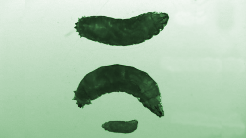
These are three Drosophila larvae that are all the same age. In the left panel, the larva at the top (npc1/+) carries only a single defective copy of the npc1 gene and grew at a normal rate to the 3rd instar stage. The larva at the bottom is defective in both of its npc1 genes (npc1/npc1), failed to grow, and stayed at the 1st instar stage. I’ll describe the larva in the middle below. The right panel illustrates how we distinguish npc1/+ from npc1/npc1 larvae. The npc1/+ larvae express the green fluorescent protein strongly in a portion of the gut (top), while npc1/npc1 larvae do not (middle and bottom).
Why don’t npc1 larvae mature? Since most npc1 mutants do not develop beyond the 1st instar stage, Huang and colleagues tested whether they are deficient for 20E, the steroid hormone that promotes larval development. That experiment was conducted by adding 20E to the larvaes’ food. Npc1 larvae survive to later stages with this extra 20E. Taking that result a bit further, feeding the larvae chemical precursors of 20E, cholesterol or 7-dehydrocholesterol, also allow npc1 mutant larvae to survive to later developmental stages. We have confirmed those results with our npc1 flies. The middle larva in the image above is homozygous for the npc1 gene. It was fed 7-dehydrocholesterol and developed normally to the 3rd instar stage.
Like human cells defective for npc1, Huang et al. showed that the npc1 mutant flies have accumulation of cholesterol in abnormal intracellular organelles. This “trapping” of cholesterol might be a big problem. In healthy cells, cholesterol travels within the cell to become a substrate in steroid hormone synthesis at defined cellular locations. The authors proposed a model that flies with defective Npc1 proteins are 20E deficient because much of their cholesterol is trapped and therefore unavailable to be a substrate in 20E synthesis. That model is supported by their experiments showing that an increase in dietary amounts of 20E precursors, 7-dehydrocholesterol and cholesterol, allowed npc1 mutants to survive to more advanced developmental stages.
Might cholesterol deficiency explain some aspects of the human disease? Huang and colleagues nicely make an argument to support that idea when stating that the irregular sterol accumulation in organelles of npc1 mutant flies and mammalian cells indicates a common sterol trafficking function that has existed for at least half a billion years. In fact, mice with npc1 mutations are testosterone deficient (Roff et al., 1993) and have decreased levels of steroid hormones in the brain (Griffin et al., 2004). Brain steroids are known to promote neuronal survival. Perhaps the neurodegeneration observed in Niemann-Pick type C animal models and patients is explained by a neurosteroid deficiency.
References
Huang, X. et al. A Drosophila model of the Niemann-Pick type C lysosome storage disease: dnpc1a is required for molting and sterol homeostasis. Development 132: 5115-5124 (2005).
Fluegel, M.L. et al. Mutations of a Drosophila NPC1 gene confer sterol and ecdysone metabolic defects. Genetics 172: 185-196 (2006).
Roff, C.F. et al. The murine Niemann-Pick type C lesion affects testosterone production. Endocrinology 133: 2913-2923 (1993).
Griffin, L. D. et al. Niemann-Pick type C disease involves disrupted neurosteroidogenesis and responds to allopregnanolone. Nat. Med. 10: 704-711 (2004).
