Niemann-Pick Type A (NPA) affects about 1 in 250,000 individuals, with a higher frequency in individuals of Ashkenazi Jewish descent, where it’s about 1 in 40,000. NPA is caused by a mutation in the SMPD1 gene, a gene encoding acid sphingomyelinase which, when mutated, stops the conversion of sphingomyelin into ceramide. This causes an accumulation of sphingomyelin in the cell which, over time, can lead to tissue and organ failure. Perlara is collaborating with Wylder Nation on the NPA PerlQuest to find possible treatments. This post lays out how we went about designing an NPA fibroblast screen.
3 parts to a PerlQuest
PerlQuests have three major parts. The first, identifying a disease phenotype/screenotype for drug screening across model organisms: C. elegans, Drosophila, human cells or yeast. The second, running the screen with a 2,560 repurposed compound library, and finally with our larger 20,000-50,000 compound library, to find novel compounds. When we find a promising hit in either library, we follow up by characterizing the mechanisms of action (MOA) for the compound. We want to understand what biological pathways the compound is impacting and how the compound might scale to humans.
While doing some MOA experiments on a novel compound we had previously identified from a of a C. elegans screen for Niemann-Pick Type C (NPC), we identified a possible screenotype for a related disease, NPA. We found that by using a fluorescent sphingomyelin marker, BODIPY-C5 sphingomyelin, we could visually see a significant difference between patient NPA fibroblasts and wild type cells.

Figure 1 – Accumulation of C5 sphingomyelin in NPA patient fibroblasts
Counting the puncta
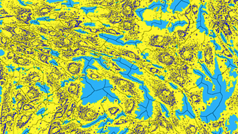
Though we could outwardly observe a huge distinction between patient NPA fibroblasts and wild type cells, simply visualizing a difference is not sufficient for a screen, which compares thousands of wells over many 384-well plates. It’s too subjective and time-consuming. Therefore, we created a data analysis tool using Custom Module Editor which could identify the individual puncta – pinpoints of high fluorescence – and count them. That count of total puncta could then be divided by the number of cells in a well giving us a total puncta per cell reading (Figure 2). Comparing this readout with our controls pulls out hits from the screen, compounds that reduce the accumulation of sphingomyelin in the cells.

Figure 2 – Result of C5 Sphingomyelin cell mask
Designing and running the NPA fibroblast screen
Armed with a screenotype and cell mask we ran trial plates to confirm the strength of the screenotype. We saw the same visual difference between our two controls, wild type, and NPA fibroblast in the presence of DMSO, and saw the total puncta-per-cell reading reflected in the mask (Figure 3).
After confirming we had good controls, we moved on to designing the NPA fibroblast screen. We wanted to use the fluorescent marker after incubation of compounds with the patient cells to ensure that the compounds were clearing the sphingomyelin, not just blocking the uptake of the fluorescent marker. Our final design was to first stain the cells with the fluorescent marker, then incubate the fibroblasts in compound for 48 hours, and finally to use the mask to see if any compounds reduced the sphingomyelin levels back to wild type.

Figure 3 – Puncta-per-cell reading reflected in the mask
Knowing we had good controls we moved ahead and ran a trial library plate. When running a trial plate with unknown compounds, the best-case scenario is to see all possible extremes: compounds that increase puncta, decrease puncta, and toxic compounds. These extremes show us that the screen is sensitive to compound interactions, and is viable for large scale screening. We saw all the desired scenarios (Figure 4) and confirmed that our controls maintained their separation giving us the “green light” to move forward with the screen.

Figure 4 – Results from our trial plates
We ran the screen following the same experimental design as the trial plate. Patient cells were stained with the probe, incubated for 48 hours in compound, and lastly imaged under a fluorescent light. The images were then processed through the above discussed mask and wells with puncta numbers similar to the wild type were visually inspected and pulled as hits. We ran three replicates of the screen to insure accuracy and compared the hit/toxic wells across all three. We ended up with 5 strong hits across all three replicates.
What’s next?
The next steps for us are to do a dose curve on those compounds to see if they can replicate their impact on the cells at lower concentrations (making them better candidates for humans), and to characterize their mechanisms of actions, giving us a better understanding of how they are reducing sphingomyelin levels. If the hit compounds show promise, we can move on to more animal models and, hopefully, into the hands of patients. Stay tuned!
