An update on the development of PMM2 Drosophila models, and our work to design high throughput drug screen for PMM2 deficiency —
Phosphomannomutase 2 deficiency (PMM2-CDG) is the most common type of congenital disorder of glycosylation resulting from a defect in synthesis of oligosaccharides, or glycans, that are attached to asparagine residues of many proteins in cells by a process called N-glycosylation. The PMM2 gene encodes an enzyme that catalyzes the conversion of mannose-6-phosphate (M6P) to mannose-1-phosphate (M1P), the first committed step of N-glycosylation. Mutations in PMM2 either reduce or eliminate the activity of the enzyme and lead to variable clinical features including developmental delay, muscle weakness, abnormal fat distribution, mild cognitive disability, inability to walk independently, seizures, liver and heart dysfunctions in PMM2 patients (Freeze et al., 2015; Jaeken and Matthijs, 2007). Recently my colleagues at Perlara, Jessica Lao and Sangeetha Iyer, posted on yeast and worm models of PMM2. This post is an update on our development of fly models of PMM2 and our assay optimization for a high-throughput drug screen for compounds that rescue PMM2 deficiency in flies.
In Stage 1 of our PerlQuest partnership with Maggie’s Cure, we aimed to develop new fly models of PMM2 by generating null and hypomorphic alleles of the fly PMM2 ortholog (CG10688, pmm2), and adapt disease-relevant phenotypes of these PMM2 mutants for high-throughput screens. PMM2 is an evolutionarily conserved gene, which shares 56% protein identity to the fly PMM2 overall and 100% identity to the active sites involved in substrate (M6P) binding and conversion to product (M1P). This high conservation increases the likelihood of discovering potential drugs effective against PMM2-CDG from whole-organism drug screens.
Past PMM2 Drosophila models
Two years ago Kendal Broadie and colleagues published a fly model of PMM2 to study the biology of PMM2 deficiency (Parkinson et al., 2016). They generated pmm2 null alleles (pmm2FS1 and pmm2FS2) by introducing frame shift mutations (FS1 and FS2) in the coding region of pmm2, which lead to truncated PMM2 proteins upon translation. They found that homozygous loss-of-function alleles pmm2FS1 and pmm2FS2 are lethal during development. Using pmm2 alleles and RNAi mediated targeted loss of function of pmm2 in whole body or in specific tissues, they demonstrated that PMM2 plays roles in regulation of lifespan and coordinated movements (Parkinson et al., 2016). Moreover, their studies in the nervous system of flies revealed that lower PMM2 activity increases synaptic excitability and also leads to more synaptic boutons and neuronal branching compared to controls, indicating a critical role of PMM2 in regulation of synaptogenesis and pruning of neuronal processes. They found that PMM2 positively regulates both extracellular matrix proteinase and trans-synaptic signaling pathways to regulate synaptic growth in the nervous system.
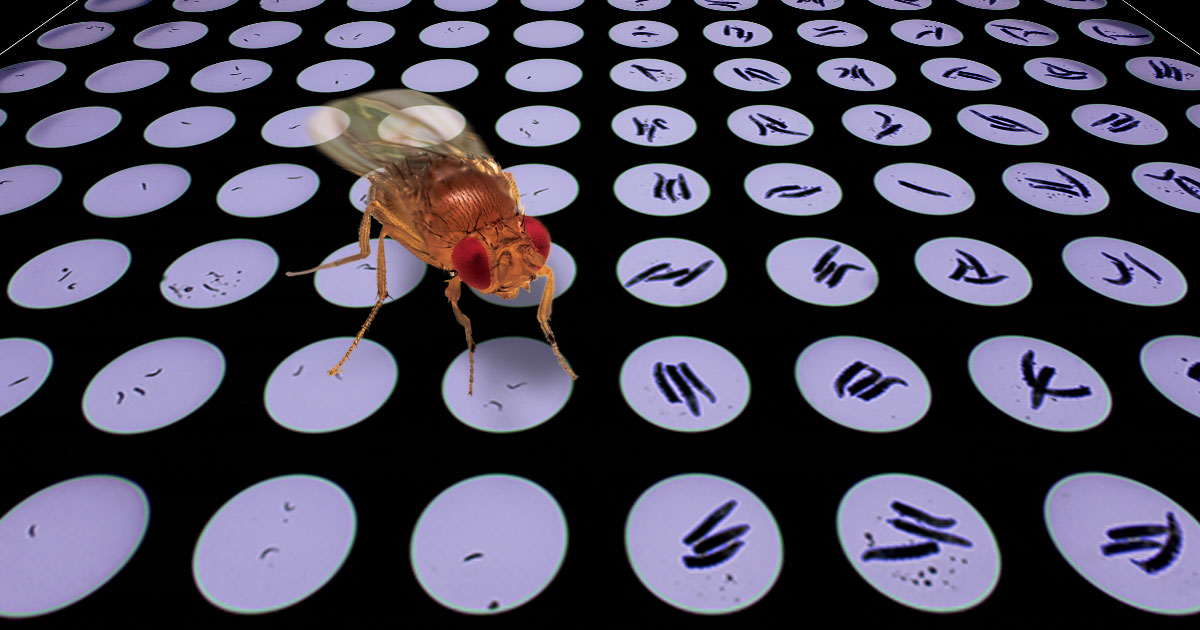
Generating Perlara pmm2 fly mutants
We independently generated our own null pmm2 alleles using the CRISPR/Cas9 gene editing technique. Using guide RNAs designed around the start and stop codons of pmm2 coding region, we replaced the protein coding sequence of PMM2 with a red fluorescent protein (dsRED) or a green fluorescent protein (GFP) (Figure 1). Initial characterization of these pmm2 alleles, hereafter referred to as pmm2dsRED, pmm2GFP#1, pmm2GFP#3, showed that complete loss of function of pmm2 is lethal (consistent with Broadie lab alleles), demonstrating the essential function of PMM2 enzyme in survival of the organism during development.

Figure 1: Creating Perlara PBC Drosophila pmm2 alleles using the CRISPR/Cas9 homology dependent DNA repair system
Characterizing lethality of pmm2 null larvae
Next we asked why and when homozygous pmm2 null alleles are lethal during development. To address these questions, we designed experiments to monitor the growth and survival of pmm2 null individuals during their embryonic and larval stages (Figure 2). We found that homozygous pmm2 null larvae are much smaller in size at 2nd and 3rd larval instar stages compared to heterozygous pmm2 null larvae. We compared the growth phenotype of homozygous pmm2dsRED, pmm2GFP#1, pmm2GFP#3 larvae to the Broadie lab lines (pmm2FS1 and pmm2FS2) and found that all null pmm2 alleles had very similar larval growth defect (Figure 2).
These findings raised another question: Did homozygous pmm2 null larvae progress through development at the same rate as heterozygous control larvae but were stunted in size, or are the homozygotes developmentally arrested? To answer this question, we dissected the larval mouth hooks from homozygous and heterozygous pmm2dsRED and pmm2FS1 larvae at the 3rd larval instar stage. The mouth hooks from heterozygous pmm2dsRED or pmm2FS1 larvae developed normally whereas the morphology of the mouth hooks from the homozygous pmm2dsRED and pmm2FS1 looked similar to those of younger larvae (Figure 2) indicating that homozygous pmm2 null larvae had a larval growth arrest between 1st and 2nd larval stages.
Since all pmm2 alleles behaved very similarly in our larval assays in our hand, we examined the time of lethality using pmm2FS1 allele in petri dishes (since our pmm2 alleles were not sufficiently expanded at the time of these experiments). We found that 44.7% of the homozygous pmm2FS1 larvae survived four days and none survived five days after egg laying as reported previously (Parkinson et al., 2016). Because of the high lethality rate of homozygous pmm2 null allele, we decided to visualize the larvae two days after they are being exposed to drug media.

Figure 2: Comparing larval growth phenotypes of Perlara and Broadie laboratory pmm2 null alleles (A) Third instar larvae homozygous or heterozygous for pmm2dsRED, pmm2GFP#1, pmm2GFP#3 (Perlara alleles), or pmm2FS1, or pmm2FS2 [Genotypes w;;pmm2dsRED/ pmm2dsRED or w;;pmm2dsRED/ TM3SerGFP (panel 1), w;;pmm2GFP#1/ pmm2GFP#1 or w;;pmm2GFP#1/ TM3SerGFP (panel 2), w;;pmm2GFP#3/ pmm2GFP#3 or w;;pmm2GFP#3/ TM3SerGFP (panel 3), w;;pmm2FS1/ pmm2FS1 or w;;pmm2FS1/ TM3SerGFP (panel 4), and w;;pmm2FS2/ pmm2FS2 or w;;pmm2FS1/ TM6B, Tb (panel 5)]
(B) Larval mouth hooks from heterozygous and homozygous pmm2dsRED and pmm2FS1 larvae at the 3rd larval instar stage [Genotypes w;;pmm2dsRED/ pmm2dsRED (panel 2 and 3) or w;;pmm2dsRED/ TM3SerGFP (panel 1) and w;;pmm2FS1/ pmm2FS1(panel 5 and 6) or w;;pmm2FS1/ TM3SerGFP (panel 4) and schematic representation of expected larval mouth hooks at the 1st, 2nd and 3rd larval instar stages
Optimizing conditions for a high-throughput screen
Our earlier studies show that some fly disease models can be sensitive to DMSO, the solvent that we use to dissolve the compounds in our chemical libraries (Rodriguez et al., 2018). To test if pmm2 null allele is sensitive to DMSO, we treated homozygous and heterozygous pmmFS1 larvae with various concentration of DMSO (0.05%, 0.1%, 0.2%, 0.3%, 0.4% and 0.5%) for two days in 96-well plates and found that homozygous and heterozygous pmm2FS1 larvae equally well tolerated DMSO at the concentrations tested. We also verified these DMSO results in petri dish experiments by monitoring both larval size and survival.
Using 0.1% DMSO concentration, we then tested a few compounds (20 Ecdysone, ecdysone, and Perl101) that were previously identified in our drug screens as potent suppressor of other disease models. As expected, none of these compounds had an effect on the size or survival of pmm2 homozygous larvae in 96-well plates (Figure 3) and petri dish experiments.
Because of the early lethality of homozygous pmm2 null larvae, we also considered the possibility of using heterozygous pmm2 null larvae in combination with a chemical modifier. We tested if pmm2 heterozygotes have altered sensitivity to bortezomib, a proteasome inhibitor that also causes ER stress. We found that at 10µM, 12µM and 14µM Bortezomib concentrations, the size of pmm2 null larvae was significantly different than wild-type larvae (Figure 4). To our surprise, heterozygous pmm2 larvae were less sensitive to bortezomib treatment than wild-type. This result is the opposite to what we found with homozygous PMM2 mutant worms, which are more sensitive to bortezomib than controls. Therefore, we decided to use homozygous pmm2 null for a drug repurposing screen. Currently we are in the process of preparing our pmm2dsRED null allele and pmm2 deletion lines generated by CRE recombinase from our pmm2GFP#1, pmm2GFP#3 alleles for the screen. Our plan is to use 8-10 larvae per well in 96-well plate and see if any of the compounds in our Microsource library can rescue larval growth arrest and lethality phenotypes of pmm2 null mutant in a 3-day assay.

Figure 3: Testing effects of select compounds on homozygous and heterozygous pmm2FS1 in 96-well plate in a 2-day assay

Figure 4: Testing effects of proteasome inhibitor bortezomib on heterozygous pmm2FS1 and wild type larvae in 3-day assay. (A) Images of third instar larvae treated with 10-18mM concentration of bortezomib. (B) Quantification of larval size via image J. **** P < 0.001, *** P= 0.006 Genotypes w;;pmm2FS1/ TM3SerGFP (pmm2 heterozygous) or w1118
Learn more about PMM2-CDG PerlQuest
References
1- Freeze, H. H., Eklund, E. A., Ng, B. G. and Patterson, M. C. (2015) Neurological aspects of human glycosylation disorders. Annu. Rev. Neurosci. 38, 105-125.
2- Jaeken,J. and Matthijs, G. (2007) Congenital disorders of glycosylation: a rapidly expanding disease family. Annu. Rev. Genomics Human Genet. 8, 261-278.
3- Parkinson, V.M., Dookwah M., Dear M.L., Gatto, C.L., Aoki, K., Tiemeyer, M., Broadie, K. (2016) Synaptic roles for phosphomannomutase type 2 in a new Drosophila congenital disorder of glycosylation disease model. Dis. Model Mech. 9(5), 513-527.
4-Rodriguez, T.P., Mast, J.D., Hartl, T., Lee, T., Sand, P., Perlstein, E.O. (2018) Defects in the Neuroendocrine Axis Contribute to Global Development Delay in a Drosophila Model of NGLY1 Deficiency. G3.300578
