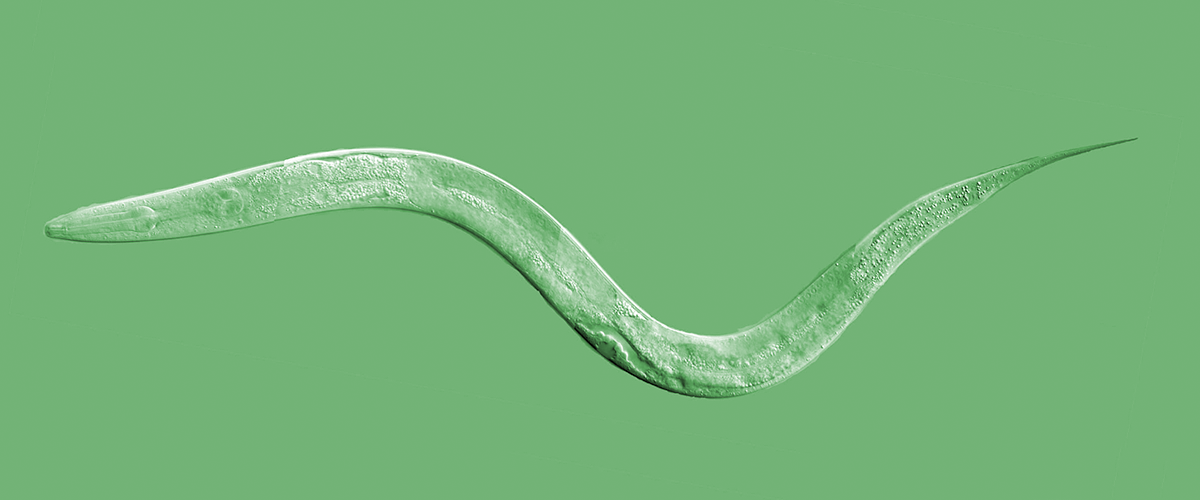
In 2016, Perlara and Wylder Nation launched a PerlQuest to find a cure for Niemann-Pick Type A (NPA). We have also been developing simple organism disease models for Niemann-Pick Type C (NPC) as a part of our research collaboration with Novartis. An interesting extension to our work on NPC and NPA disorders is the possibility that we may be able to reuse compounds in the treatment of another lysosomal storage disorder, Gaucher disease (or GD). We have been piloting work with a mutant strain of C. elegans to see what possibilities our pipeline offers for this disease.
An intro to Gaucher disease
GD is a relatively common autosomal recessive disorder with an incidence of about 1 in 100,000. It is the most common inherited disorder amongst the Ashkenazi Jewish population. Recent studies have also shed light on a relationship between the causative gene for GD and an increased risk for Parkinson’s, making any treatment for this condition a very exciting possibility.
GD is caused by a mutation in the GBA1 gene, completely eliminating or severely disrupting the ability for cells, particularly macrophages in humans, to use the enzyme beta-glucocerebrosidase (GCase) to break down glucocerebroside (and related substances). These products subsequently build up in cells, causing a “crinkled paper” appearance on histology (also called Gaucher Cells).
GD is a spectrum disorder, probably in relation to the extent of dysfunction caused by particular mutations. It is usually clinically classified on a three tier system (GD-1, 2, & 3). In most (human) cases of all three types, cells in the spleen and liver are progressively affected. In more severe cases (GD-2 &3), the nervous system is also affected, often leading to premature death.
What about Gaucher worms?
How would we go about modeling this in worms, which do not have spleens or livers (or even proper macrophages!)? Adding to these challenges, worms are also extraordinarily hard to perform histology on in part due to their dense outer coating known as the cuticle. This makes staining of intact morphological structures challenging, to say the least. We need a “screenotype” or some phenotypic manifestation of the mutation and related deficits that is easily detectable en masse. By that metric, we can see if compounds in our screens rescue the deficit.
Worms have four copies (or orthologs) of the human GBA1 gene (gba-1, 2, 3, & 4), each with varying degrees of similarity to human GBA1. Of the 4 orthologs, gba-3 and gba-4 are hypothesized to possess lysosomal glucocerebrosidase activity. Thus, we decided to focus on gba-3 and gba-4. Luckily, a gba3-null mutant, VC3135, was available from the Caenorhabditis Genetics Center (CGC).
We first embarked on phenotyping this available mutant. Using a biochemical assay, we determined that gba-3 null worms showed reduced glucosylceramidase activity, much like humans with GD. Unluckily, even without this gene, the worms demonstrated no overt phenotype, i.e., they live out their lives (from all we can tell) just the same as worms with fully formed copies of all four gba orthologs. They show no gross abnormalities during development or in behavior.
To find a screenotype!
Using quantitative PCR, we found that while levels of gba-3 were reduced to approximately 50% in age-synchronized day 1 adult gba-3 null worms (VC3135) relative to wildtypes (N2), they were not completely eliminated. The levels of the other orthologs were also not significantly changed in the gba-3 null worms relative to wildtypes suggesting no major compensation from any of the other genes. This suggested that residual gba-3 and possibly gba-4 were likely responsible for the observed enzyme activity in the worms. While enzyme activity is reduced, no overt phenotypes were noted in the gba-3 null animals.

GBA-3 null (VC3135) worms have approximately 50% enzyme activity relative to wildtype worms (N2)
We therefore asked if we could knock down gba-3 further to induce a phenotype. We set about to induce a phenotype by inhibiting gba transcripts through a method known as RNA-interference, or RNAi. Worms are particularly suited to this methodology, as double-stranded antisense RNA can be inserted directly into the bacteria worms feed upon. We attempted knocking down gba3 and gba4. Unfortunately, in all conditions, the worms showed the same happy sinusoidal movement as their control-condition peers. We saw no easily screenable metric; no overt changes size, motility, feeding, swimming, life expectancy, or reproduction. Further transcript quantitation from these studies is underway.
Next, we attempted a drug interference to exacerbate the phenotype. Conduritol B epoxide (CBE) is an irreversible inhibitor of glucocerebrosidase. We introduced fifteen L1 larvae of a gba-3 null mutants into wells of a 384-well plate containing concentrations from 500nM to 50uM CBE. Two columns of control wells were introduced on either end with 137.5 and 250nL’s of DMSO, representing respectively the average and upper bounds of total DMSO in experimental wells. All plates were exposed for 6 days with shaking at 20C.
 |
 |
 |
| From left to right: Representative images of DMSO control, 500nM CBE and 50µM CBE after 6 days of exposure. | ||
 |
||
| Boxplot of area occupied by worms (arbitrary units) at different concentrations of CBE | ||
At the end of the exposure, we imaged the wells and used image processing algorithms to extract the areas occupied by worms per well. As can be seen from the figures and example images above, no overt change was seen in the experiment.
Because we have the advantage of studying diseases in parallel model organisms, we decided to take another stab by following our fly teams example and attempted a heat-shock experiment to see if the gba-3 null worm response was different from the wild-type. We tested the effect of heat shock on gba-3 null worms and N2s under many conditions. Like our GBA1a and GBA1b null flies, gba-3 null worms are more sensitive to heat shock-induced paralysis compared to wildtype. When exposed to heat for short periods of time, gba-3 null worms did not move as much.
A change was seen when the worms were incubated from 30 minutes to one hour at 37C! Wildtype worms were less sensitive to the heat and recovered faster. The change was qualitatively obvious to our observers; however, developing a quantifiable metric remains challenging. We are exploring new imaging options and automated analyses to see how best to detect these changes.
 |
 |
Left panel: Recovery plot of percent animals (WT and GBA-3 nulls) moving post 30 minutes of exposure to 30ᵒC or 37ᵒC. Right panel: Recovery plot of percent animals (WT and GBA-3 nulls) moving post 60 minutes of exposure to 30ᵒC or 37ᵒC.
Moving forward
As we finish the analysis we have begun and continue assay development, we hope to find a screenotype which can allow us to discover candidate compounds against this model of Gaucher Disease. The applications of any drug which treats this debilitating condition are apparent and given the prevalence and likely connection to Parkinson’s Disease, crucial to quality of life for thousands of people throughout the world.
Photo credit: Modified from the original by Judith Kimble, University of Wisconsin


